My Account Details

ISBN10: 0071637974 | ISBN13: 9780071637978

Step 1 . Download Adobe Digital Editions to your PC or Mac desktop/laptop.
Step 2. Register and authorize your Adobe ID (optional). To access your eBook on multiple devices, first create an Adobe ID at account.adobe.com. Then, open Adobe Digital Editions, go to the Help menu, and select "Authorize Computer" to link your Adobe ID.
Step 3. Open Your eBook. Use Adobe Digital Editions to open the file. If the eBook doesn’t open, contact customer service for assistance.
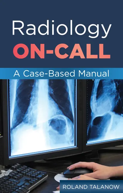
Publisher's Note: Products purchased from Third Party sellers are not guaranteed by the publisher for quality, authenticity, or access to any online entitlements included with the product. 200 of the most common cases for radiology on-call/emergency situations—inone uncommon guide Radiology On-Call covers the full spectrum of clinical scenarios that you are likely to see in the emergency department or during an in-house call. Two hundred cases are logically arranged by organ system, supported by 375 precise, state-of-the-art radiographs, CT, MRI, nuclear medicine and ultrasound images that accelerate on-the-spot clinical decision-making. Radiology On-Call has an easy-to-navigate, streamlined style that features annotated images and minimal text. The author provides only those facts and brief descriptions that are needed to become familiar with each entity. Features: The complete on-call radiology sourcebook, designed to help residents ensure the accuracy of radiologic interpretations, become familiar with emergency findings, and reduce on-call errors 200 highly instructive cases containing 375 radiographs, CT, MRI, nuclear medicine, and ultrasound images, many in full color Consistent organization: image, diagnosis, comments, cross-reference to online tutorial Cross-reference to interactive online tutorial: Cases are linked to an online tutorial (www.oncallradiology.com) providing many cases in a unique interactive way almost as seen on a real workstation (scroll, window, level, magnify, pan). Content intuitively organized by organ system: Chest, Abdomen, Neuro, Musculoskeletal Section-opening anatomical overviews, featuring clearly labeled radiographs, provide a solid base of knowledge for understanding subsequent material on imaging and image-guided situations Large collection of references, including links to free, open-access high-quality review articles about specific topics discussed in the book
I. Chest
1. Anatomy
2. Technique
3. Lung & Airways
4. Pleural Space
5. Mediastinum, Heart & Vessels
6. Chest Wall & Diaphragm
7. Devices
8. References
II. Abdomen
1. Anatomy
2. Technique
3. Liver & Gallbladder
4. Pancreas
5. Spleen
6. Adrenal Glands
7. Kidneys & Urinary Tract
8. Gastrointestinal Tract
9. Female Reproductive System
10. Male Reproductive System
11. Vascular System
12. Devices
13. References
III. Neuro
1. Anatomy
2. Technique
3. CT Evaluation
4. Head
5. Neck
6. Spine
7. References
IV: Musculoskeletal
1. Technique
2. Radiograph Evaluation
3. Shoulder Girdle
4. Upper Extremities
5. Pelvis and Hip
6. Lower Extremities
7. References
References
Index
Need support? We're here to help - Get real-world support and resources every step of the way.