How 3D Models Enhance Learning: Examples of Views Using the Head & Neck 3D Model
Head and neck 3D model | Anatomy & Physiology Revealed (APR) 4.0.
- Succeed in A&P
- Article
- Blog
- Higher Education
In January 2020, a head and neck 3D model was added to Anatomy & Physiology Revealed (APR) 4.0. This 3D model is found in Module 15, Regional Anatomy, or can be accessed from other body systems (skeletal, muscular, nervous, cardiovascular, and endocrine) by using this icon. 
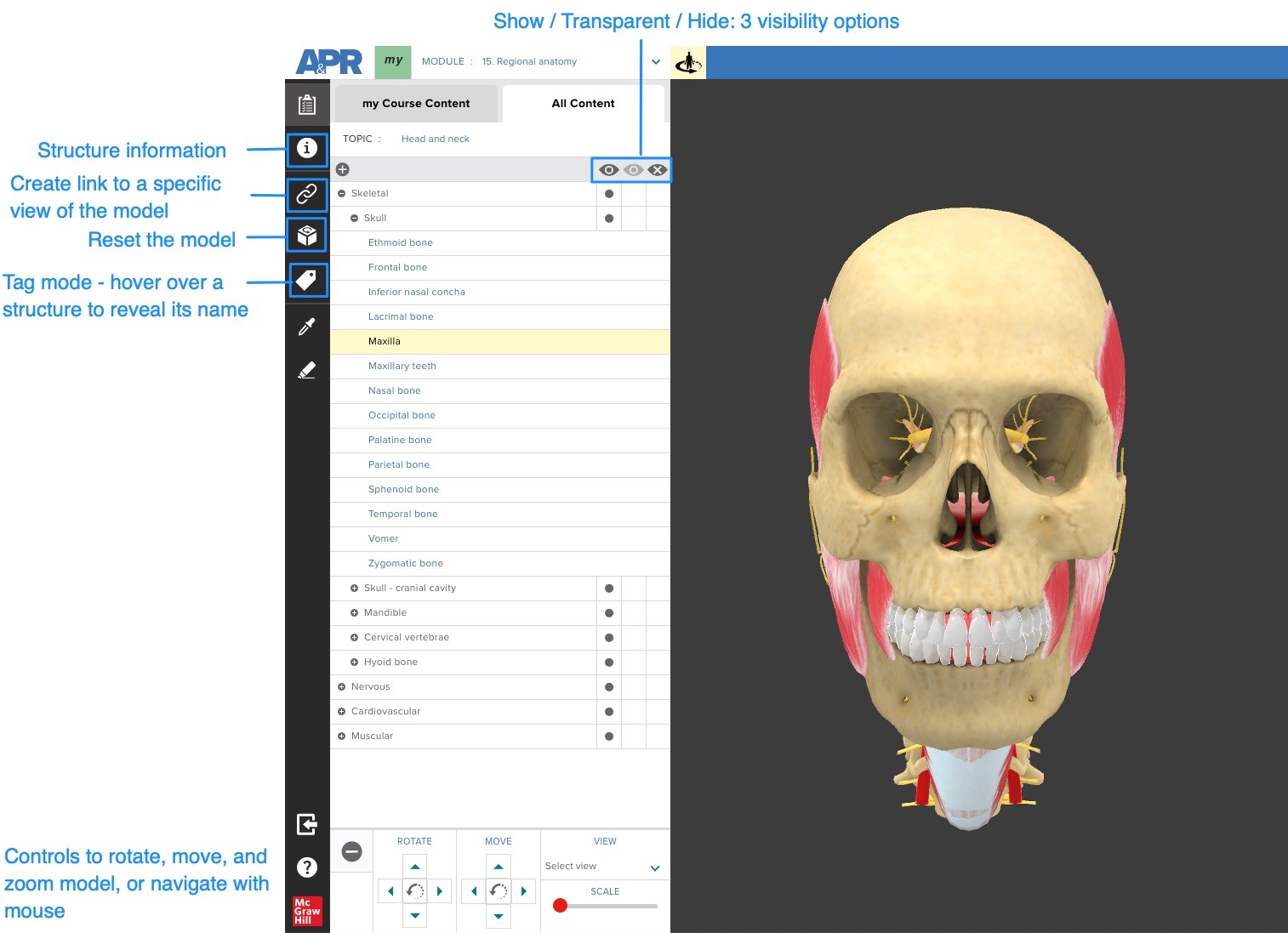
The image below highlights some of the important control icons that are used with the 3D model.
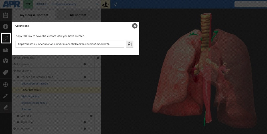
The “Create link” icon is a quick way to create a bookmark that will allow someone else to access the same exact location in APR 4.0.
How do I use this link?
Once you have the location you’d like to share, click the clipboard icon (blue arrow) to copy the link to your clipboard and then paste this link into a document, presentation slide, website, or in your LMS. Someone trying to access this link simply needs to first log into APR (since it is a password-protected website) or already have a browser tab open to APR, and then click on the link to be taken to this same exact location in APR. This feature works with the 3D model and in any other mode in APR such as dissection or histology.
How might you use this “Create link” with the 3D model in your course?
- Use the link feature to bookmark an interesting view you have created with the 3D model. You can then share this link with your students or use it to quickly access this specific view of the 3D model during lecture or lab for demonstration purposes.
- An active learning activity: scavenger hunt. Give your students a specific task with the 3D model and ask them to copy their link and send it you when they have completed the task. For example, you might tell them to find a view in the 3D model that will allow them to observe the vestibulocochlear nerve entering the internal acoustic meatus.
Below are some links to views in the head and neck 3D model I have found interesting and may help you in your teaching.
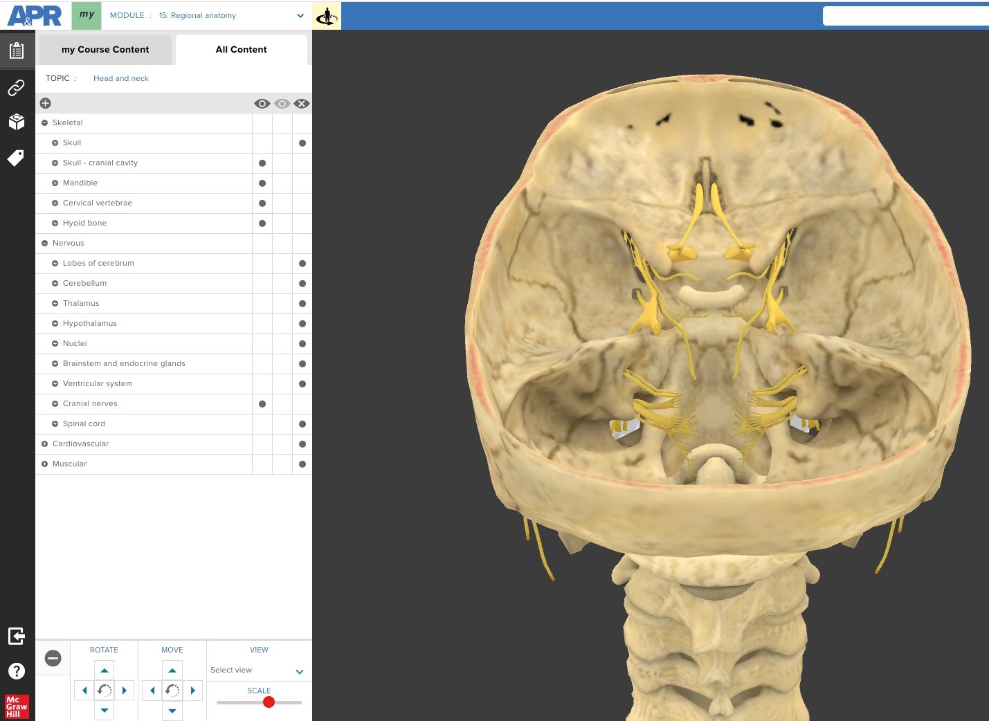
Superior view of cranial cavity
This view allows a student to observe the associated skull foramina of each pair of cranial nerves. With the 3D model, they can tilt and rotate the model so they are able to get an optimal view of each cranial nerve and associated skull foramina. This activity allows a student to replicate what they can do in a lab with a skull and pipe cleaners. http://anatomy.mheducation.com/html/apr.html?animal=human&mod=187
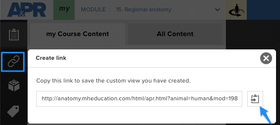
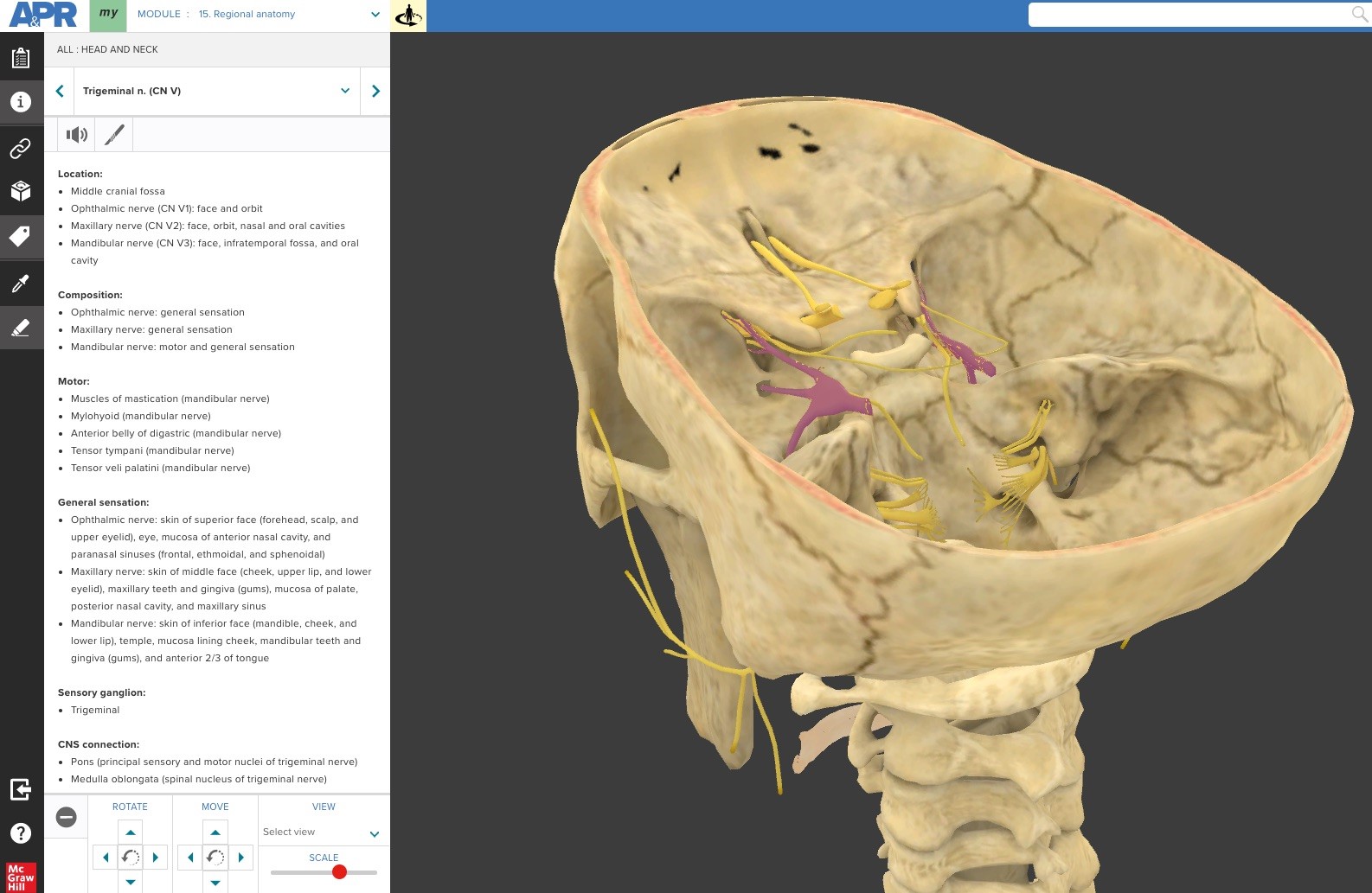
Below is the trigeminal nerve (CN V) highlighted in the 3D model using “Tag mode”. Clicking on the scalpel icon opens a window to show dissection views of the structure in the cadaveric specimen. Also available for the student is the structure information for this nerve.
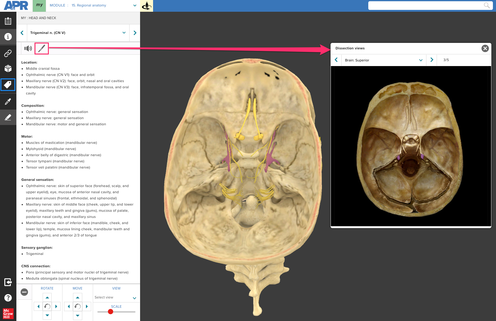
In this link, the trigeminal nerve is highlighted, and the model has been zoomed and angled to allow a student to clearly see the three different foramina associated with each division of this nerve. http://anatomy.mheducation.com/html/apr.html?animal=human&mod=199
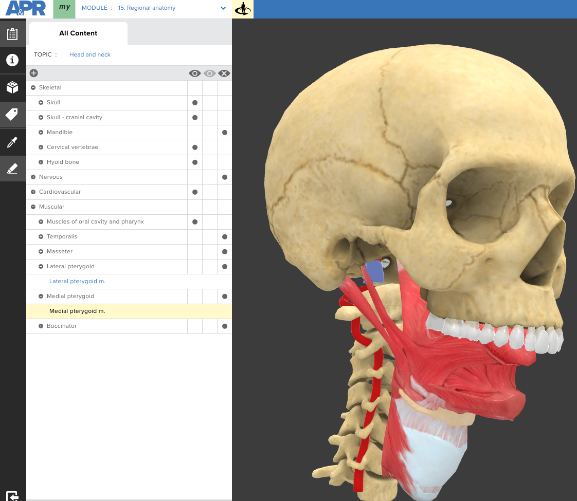
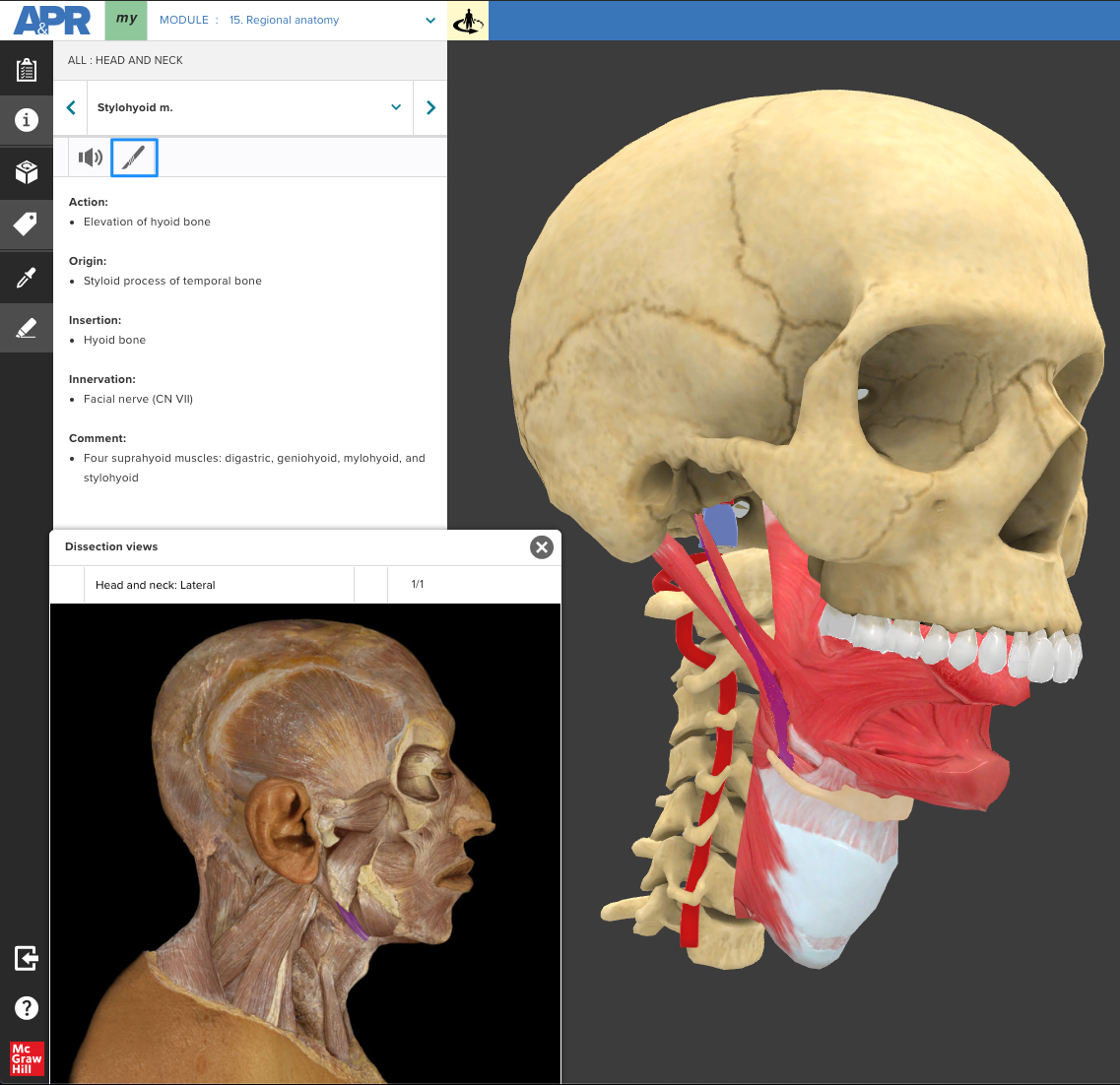
Anterior-lateral view of muscle of oral cavity and pharynx
In this view, the mandible and muscles of mastication are removed. This allows a student to explore the relationship of the suprahyoid, tongue, and pharynx muscles to one another. http://anatomy.mheducation.com/html/apr.html?animal=human&mod=18
Here is the stylohyoid muscle highlighted in the 3D model (Tag mode) and the dissection view of the stylohyoid muscle in the cadaveric specimen. Also visible is the structure information for this muscle.
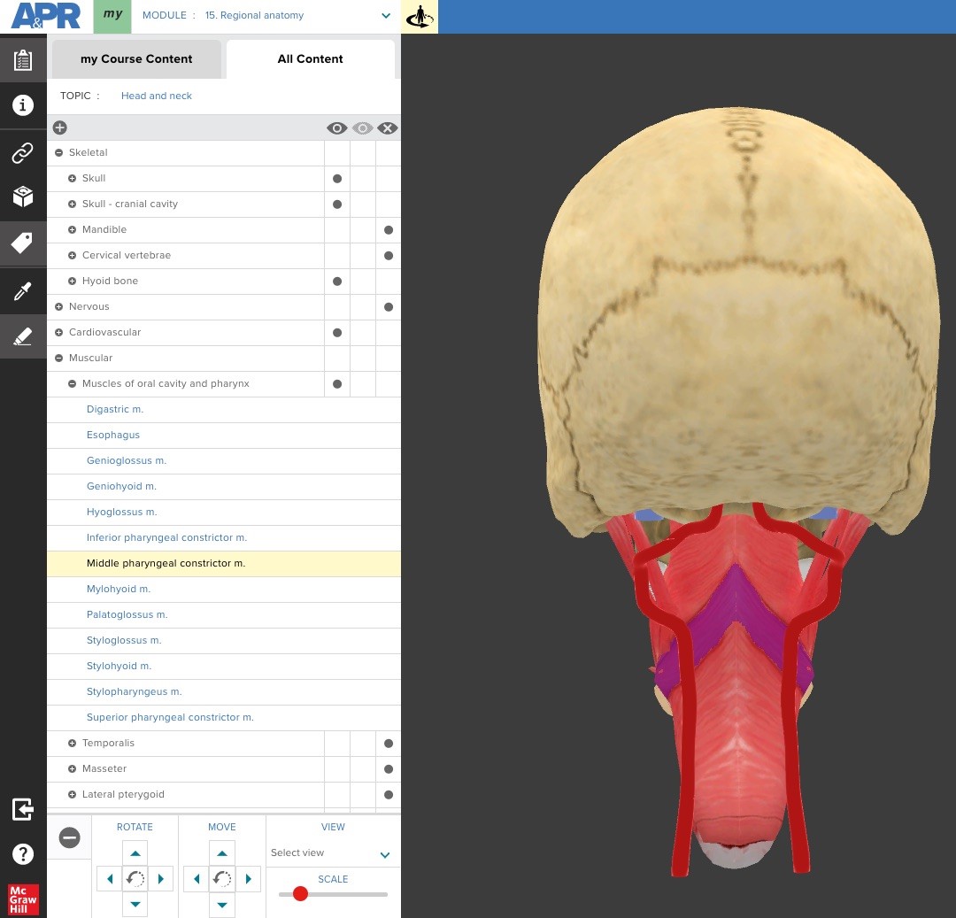
Here is a posterior view that hides the cervical vertebrae and allows a student to see the pharyngeal constrictor muscles and their relationship to one another, the hyoid, larynx, and esophagus. http://anatomy.mheducation.com/html/apr.html?animal=human&mod=190
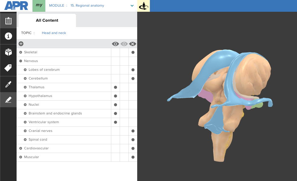
Ventricular system of the brain
This posterior-inferior view allows a student to see and explore the complex relationship of the thalamus, hypothalamus, lateral ventricles, third ventricle, fourth ventricle, pons, and midbrain to one another.
http://anatomy.mheducation.com/html/apr.html?animal=human&mod=191
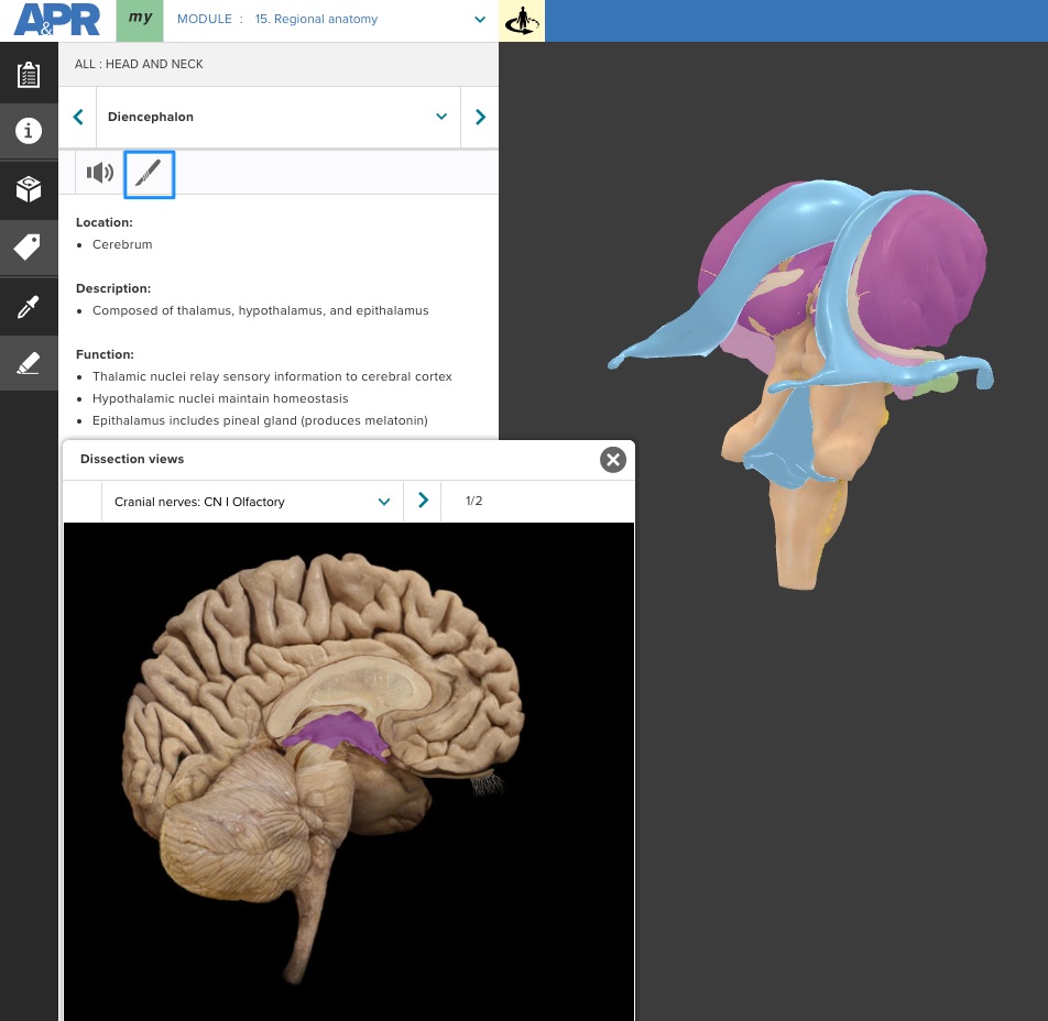
Below is the diencephalon highlighted in the 3D model and the dissection view of the structure in the cadaveric specimen. The 3D model allows a student to better visualize the shape of the diencephalon, rather than simply seeing it as a flat structure in the typical sagittal view. Also visible is the structure information.
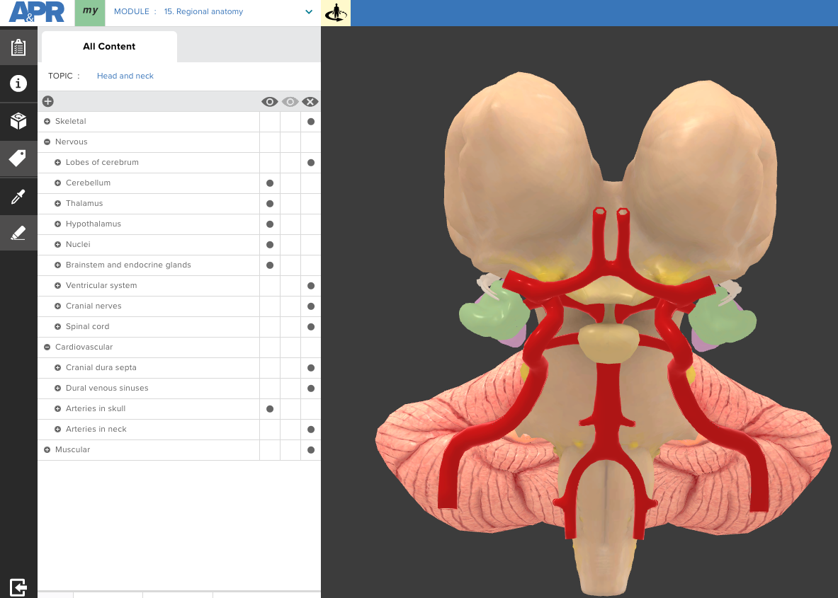
Anterior view of brainstem
In this view, a student can observe the main components of the cerebral arterial circle and its relationship to surrounding structures such as the pons, pituitary gland, and diencephalon.
http://anatomy.mheducation.com/html/apr.html?animal=human&mod=192
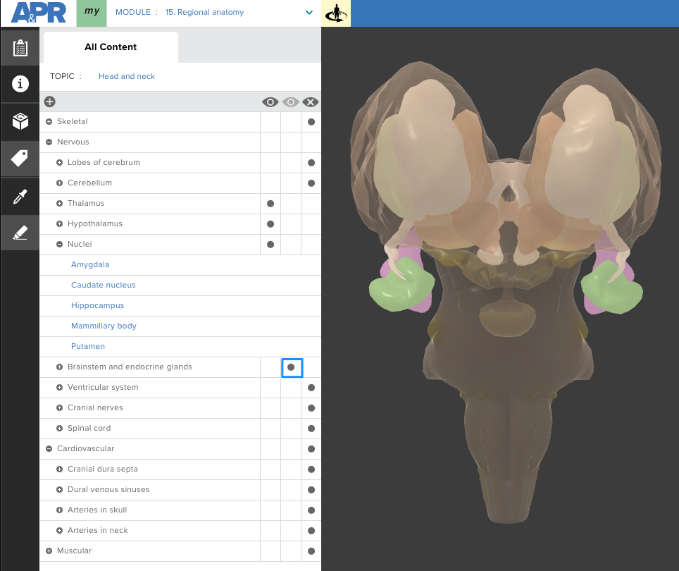
Anterior view of brainstem with transparent diencephalon
In this anterior view of the brainstem, the diencephalon and brainstem are 50% transparent. This allows the student to observe structures such as the thalamus, hypothalamus, putamen, and caudate nucleus that are not typically visible.
http://anatomy.mheducation.com/html/apr.html?animal=human&mod=194
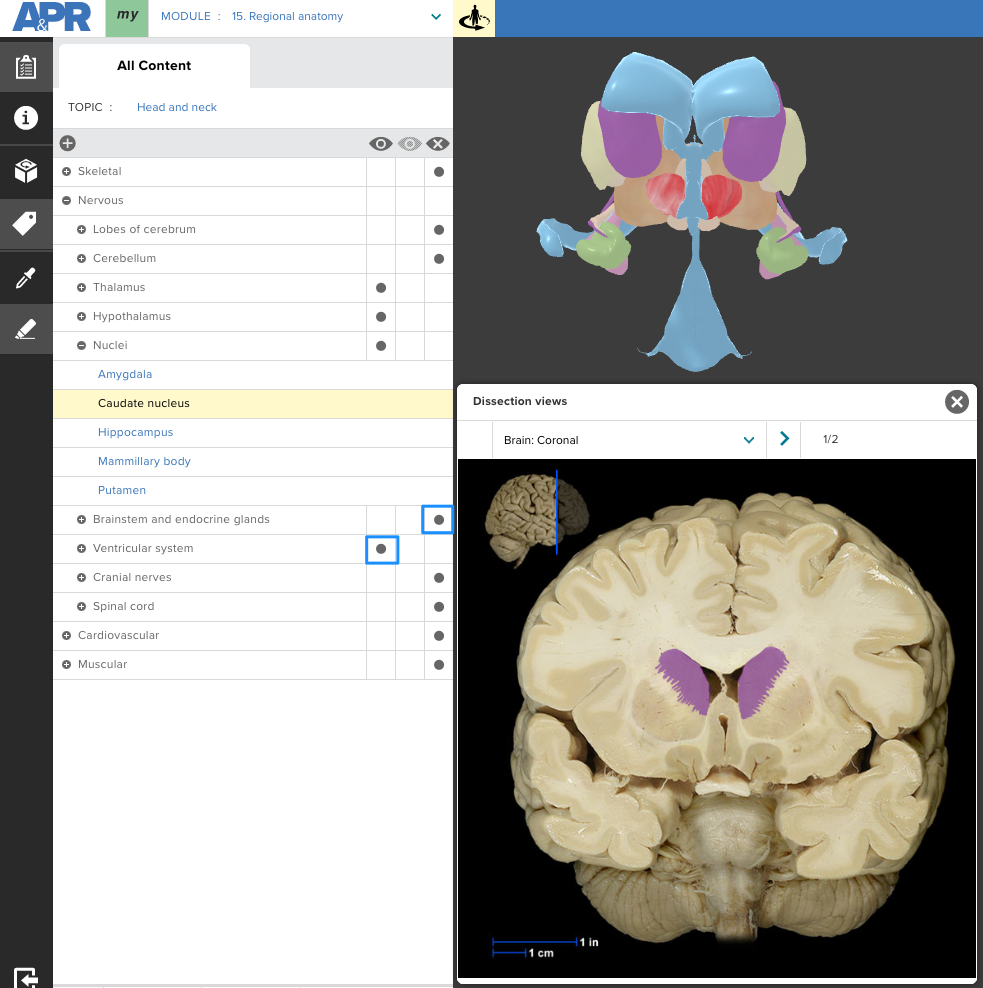
Below is the caudate nucleus highlighted in the 3D model and dissection view of the structure in the cadaveric specimen. The brainstem has been hidden and ventricular system turned on to allow a student to see the relationship between the caudate nucleus and lateral ventricles in both the 3D model and cadaveric specimen.
http://anatomy.mheducation.com/html/apr.html?animal=human&mod=195
We would love to hear from you how you have incorporated the head and neck 3D model into your course! Send us an email at A&P@mheducation.com.

Dr. Michael Koot is a Senior Director of Digital Content and Pedagogy for McGraw Hill. Before joining McGraw Hill, Michael taught human anatomy at Michigan State University where he won a campus-wide teaching with technology award. At McGraw Hill, he works at the intersection of content, pedagogy, accessibility, and technology with both internal and external stakeholders to develop effective teaching and learning tools.